Free Preparation Discussions
ARDMS AB-Abdomen Exam - Topic 4 Question 10 Discussion
Topic #: 4
Which congenital disorder is most consistent with the finding identified by the arrow on this image?
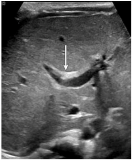
The image demonstrates a characteristic 'central dot sign' --- a hallmark finding of Caroli disease. This is best appreciated on ultrasound as a cystic dilation of the intrahepatic bile ducts with a central echogenic dot or linear structure (which corresponds to the portal vein and fibrous tissue within the dilated duct). The arrow in the image points to one such dilated duct.
Caroli disease is a rare congenital disorder characterized by segmental, saccular dilation of intrahepatic bile ducts. It is often associated with congenital hepatic fibrosis and may predispose to cholangitis, stone formation, and even cholangiocarcinoma.
Key ultrasound features of Caroli disease:
Cystic or saccular dilations of the intrahepatic bile ducts
The 'central dot sign' --- echogenic focus in the center of the dilated ducts (representing portal vein radicle or fibrous tissue)
May show associated hepatosplenomegaly or signs of portal hypertension
Differentiation from other options:
A . Sclerosing cholangitis: Typically causes diffuse or segmental biliary ductal wall thickening and stricturing; does not present with cystic dilations.
B . Alagille syndrome: A multisystem disorder often characterized by a paucity of intrahepatic bile ducts, not dilation.
D . Biliary atresia: Presents in infancy with obliteration of extrahepatic bile ducts, echogenic 'triangular cord' sign, and absence of a visible gallbladder. It does not cause ductal dilation.
Rumack CM, Wilson SR, Charboneau JW, Levine D. Diagnostic Ultrasound. 5th Edition. Elsevier, 2018. Chapter: Biliary System, pp. 152--155.
Radiopaedia.org. Caroli disease. https://radiopaedia.org/articles/caroli-disease
American College of Radiology (ACR). ACR--SPR Practice Parameter for the Performance of Pediatric Abdominal Ultrasound, 2022.
Cammy
Laura
5 days agoVivan
10 days agoEun
15 days agoCherelle
2 months agoOwen
2 months agoAnna
3 months agoLouann
3 months agoGracia
3 months agoAdell
3 months ago