Free Preparation Discussions
ARDMS AB-Abdomen Exam Questions
- Topic 1: Anatomy, Perfusion, and Function: This section of the exam measures the skills of abdominal sonographers and focuses on evaluating the physical characteristics, blood flow, and overall function of abdominal structures. Candidates must understand how to assess organs such as the liver, kidneys, pancreas, and spleen for size, shape, and movement. It also involves analyzing perfusion to determine how effectively blood circulates through these organs. The goal is to ensure accurate interpretation of both normal and abnormal functions within the abdominal cavity using sonographic imaging.
- Topic 2: Pathology, Vascular Abnormalities, Trauma, and Postoperative Anatomy: This section of the exam evaluates the abilities of diagnostic medical sonographers and covers the detection and analysis of diseases, vascular issues, trauma-related damage, and surgical alterations in abdominal anatomy. Candidates are expected to identify abnormal growths, inflammations, obstructions, or vascular irregularities that may affect abdominal organs. They must also recognize post-surgical changes and assess healing or complications through imaging. The emphasis is on correlating pathological findings with clinical data to produce precise diagnostic reports that guide further medical management.
- Topic 3: Abdominal Physics: This section of the exam measures the knowledge of ultrasound technicians in applying imaging physics principles to abdominal sonography. It includes understanding how to optimize ultrasound equipment settings for the best image quality and how to identify and correct imaging artifacts that can distort interpretation. Candidates should demonstrate technical proficiency in handling transducers, adjusting frequency, and managing depth and gain to obtain clear, diagnostic-quality images while minimizing errors caused by acoustic artifacts.
- Topic 4: Clinical Care, Practice, and Quality Assurance: This section of the exam tests the competencies of clinical ultrasound specialists and focuses on integrating patient care standards, clinical data, and procedural accuracy in abdominal imaging. It assesses the candidate ability to follow established medical guidelines, ensure correct measurements, and provide assistance during interventional or diagnostic procedures. Additionally, this domain emphasizes maintaining high-quality imaging practices and ensuring patient safety. Effective communication, adherence to protocols, and continuous quality improvement are key aspects of this section.
Free ARDMS AB-Abdomen Exam Actual Questions
Note: Premium Questions for AB-Abdomen were last updated On Jul. 09, 2026 (see below)
Which abnormality is depicted in this image of a patient who presents with a fever following a liver biopsy?
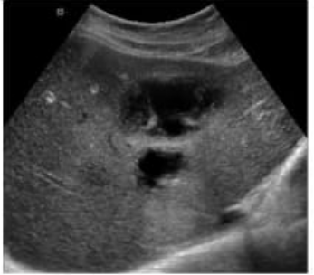
The sonographic image shows a complex fluid collection within the liver parenchyma, with internal echoes and possibly septations, consistent with an abscess. In the clinical context of post-procedural fever following a liver biopsy, a liver abscess is the most likely diagnosis.
A liver abscess appears on ultrasound as a hypoechoic or complex fluid collection that may contain internal debris, septations, or gas (which may produce reverberation artifacts). These features distinguish it from other post-procedural complications.
A cyst (Option A) typically appears as an anechoic, well-defined lesion with posterior acoustic enhancement and no internal debris---this does not match the image or clinical setting.
A biloma (Option B) is a bile collection that can appear similar to a cyst or fluid collection but typically occurs due to bile leak; however, fever and internal complexity on ultrasound more strongly suggest abscess.
A hematoma (Option D) may also appear complex but usually presents with pain and not fever unless secondarily infected. Over time, hematomas evolve in appearance but lack septations and gas unless superinfected.
Rumack, Carol M., et al. Diagnostic Ultrasound. 5th ed., Elsevier, 2018. Chapter: Hepatobiliary System, pp. 107--111.
American Institute of Ultrasound in Medicine (AIUM) Practice Parameter for the Performance of an Ultrasound Examination of the Abdomen and/or Retroperitoneum.
Which type of artifact is indicated by the arrows on this image?
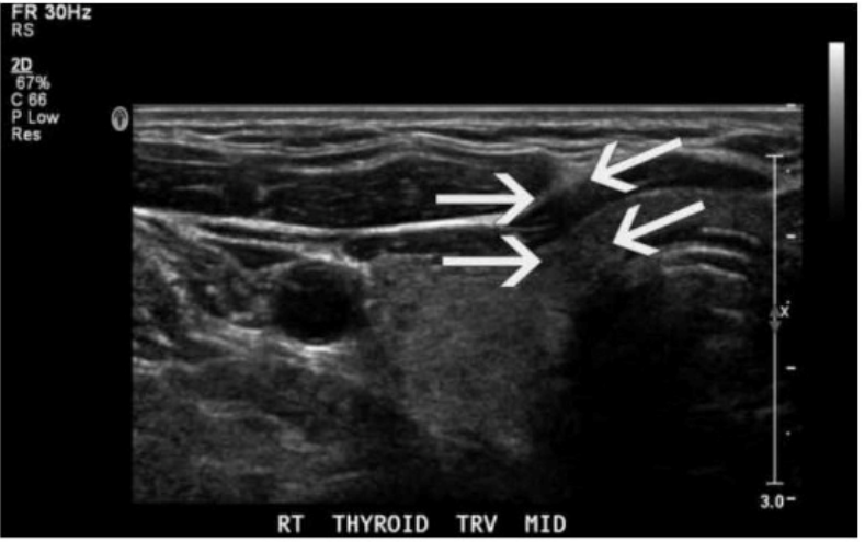
The ultrasound image of the thyroid clearly shows posterior shadowing originating from the lateral edges of a rounded structure, which is indicative of edge shadow artifact. Edge shadowing occurs when an ultrasound beam passes tangentially to a rounded or curved structure, such as a cyst or blood vessel. The difference in sound wave refraction and beam divergence at the edges leads to decreased echo signals deep to the edges, creating linear hypoechoic bands --- which is exactly what the arrows are pointing to in the image.
Edge shadow artifact is purely a result of beam physics and not a real anatomic or pathologic finding.
Key characteristics of edge shadowing:
Appears as a narrow, linear hypoechoic (dark) shadow extending deep to the edge of a curved interface (e.g., cyst, vessel, thyroid nodule)
Caused by refraction and beam deflection, leading to reduced beam intensity distal to the edges
Most commonly seen adjacent to cysts or fluid-filled structures
Differentiation from other options:
A . Focal enhancement: Appears as increased echogenicity distal to a fluid-filled structure due to lower attenuation of the sound beam through fluid (opposite of shadowing).
C . Speed error: A less common artifact that results in displacement of structures due to variation in assumed sound speed.
D . Comet tail: A reverberation artifact that appears as a series of closely spaced bright echoes, often associated with metallic objects or cholesterol crystals in adenomyomatosis.
Rumack CM, Wilson SR, Charboneau JW, Levine D. Diagnostic Ultrasound. 5th Edition. Elsevier, 2018. Chapter: Ultrasound Physics and Artifacts, pp. 38--42.
Kremkau FW. Sonography Principles and Instruments. 9th Edition. Elsevier, 2015. Chapter: Image Artifacts, pp. 132--136.
What is the most common ultrasound appearance of the pancreas in mild acute pancreatitis?
In mild acute pancreatitis, the pancreas often appears diffusely enlarged and slightly hypoechoic due to edema and inflammation. However, in very early or mild cases, the pancreas may still appear normal. Heterogeneous echotexture may develop in more severe or necrotizing pancreatitis.
According to Rumack's Diagnostic Ultrasound:
''In mild pancreatitis, the pancreas is commonly enlarged and hypoechoic due to inflammatory edema.''
Rumack CM, Wilson SR, Charboneau JW, Levine D. Diagnostic Ultrasound. 5th ed. Elsevier, 2017.
AIUM Practice Parameter for the Performance of an Ultrasound Examination of the Abdomen, 2020.
---
Which type of artifact is indicated by the arrows on this image?
The ultrasound image of the thyroid clearly shows posterior shadowing originating from the lateral edges of a rounded structure, which is indicative of edge shadow artifact. Edge shadowing occurs when an ultrasound beam passes tangentially to a rounded or curved structure, such as a cyst or blood vessel. The difference in sound wave refraction and beam divergence at the edges leads to decreased echo signals deep to the edges, creating linear hypoechoic bands --- which is exactly what the arrows are pointing to in the image.
Edge shadow artifact is purely a result of beam physics and not a real anatomic or pathologic finding.
Key characteristics of edge shadowing:
Appears as a narrow, linear hypoechoic (dark) shadow extending deep to the edge of a curved interface (e.g., cyst, vessel, thyroid nodule)
Caused by refraction and beam deflection, leading to reduced beam intensity distal to the edges
Most commonly seen adjacent to cysts or fluid-filled structures
Differentiation from other options:
A . Focal enhancement: Appears as increased echogenicity distal to a fluid-filled structure due to lower attenuation of the sound beam through fluid (opposite of shadowing).
C . Speed error: A less common artifact that results in displacement of structures due to variation in assumed sound speed.
D . Comet tail: A reverberation artifact that appears as a series of closely spaced bright echoes, often associated with metallic objects or cholesterol crystals in adenomyomatosis.
Rumack CM, Wilson SR, Charboneau JW, Levine D. Diagnostic Ultrasound. 5th Edition. Elsevier, 2018. Chapter: Ultrasound Physics and Artifacts, pp. 38--42.
Kremkau FW. Sonography Principles and Instruments. 9th Edition. Elsevier, 2015. Chapter: Image Artifacts, pp. 132--136.
Which sonographic finding is associated with normal postprocedural Doppler of a transjugular intrahepatic portosystemic shunt (TIPS)?
After successful TIPS placement, the intrahepatic portal venous branches continue to exhibit hepatopetal (toward the liver) flow, while the stent itself shows continuous, relatively high-velocity monophasic flow. Hepatofugal flow in intrahepatic branches may indicate shunt dysfunction.
According to Zwiebel's Introduction to Vascular Ultrasound:
''Normal post-TIPS Doppler shows hepatopetal flow in the intrahepatic portal veins and continuous high-velocity flow within the stent.''
Zwiebel WJ, Pellerito JS. Introduction to Vascular Ultrasound. 6th ed. Elsevier, 2019.
AIUM Practice Parameter for the Performance of Portal Venous Ultrasound, 2020.
---
- Select Question Types you want
- Set your Desired Pass Percentage
- Allocate Time (Hours : Minutes)
- Create Multiple Practice tests with Limited Questions
- Customer Support
Melissa Young
13 days agoBetty Nelson
28 days agoEdward Murphy
1 month agoAshley Garcia
2 months agoJason Edwards
2 months agoJennifer Jackson
3 months agoJessica Nguyen
2 months agoJeffrey Young
3 months agoTimothy Reed
2 months agoMichelle Thomas
2 months agoHarold Baker
2 months agoSharika
3 months agoSteffanie
4 months agoNettie
4 months agoJohnathon
4 months agoStevie
4 months agoCarey
5 months agoYoko
5 months agoMable
5 months agoNathan
5 months agoTheron
6 months agoBrendan
6 months agoFranklyn
6 months agoKris
6 months agoBuffy
7 months agoMarcelle
7 months agoMattie
7 months agoCarin
7 months agoLeonora
8 months agoShaunna
8 months agoChuck
8 months agoJohna
8 months ago