Free Preparation Discussions
ARDMS AB-Abdomen Exam - Topic 2 Question 11 Discussion
Topic #: 2
Which technique is best for demonstrating the characteristic of the small hepatic lesion identified by the arrow on this image?
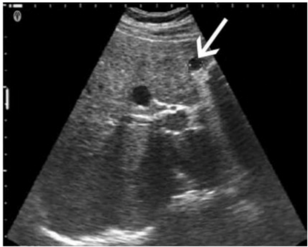
The image shows a small hepatic lesion located very close to the anterior liver capsule, as indicated by the arrow. When imaging very superficial or near-field structures like subcapsular hepatic lesions, using a standoff pad is the most effective technique for optimizing visualization.
A standoff pad (also known as an acoustic stand-off or gel pad) helps increase the distance between the transducer and the superficial target. This improves the focus and beam shape for near-field imaging and minimizes reverberation and ring-down artifacts. It allows better evaluation of superficial lesions by positioning them within the focal zone of the transducer, which is usually set a few millimeters below the probe surface.
Differentiation from other options:
A . Decrease depth: While reducing depth can help center deeper lesions in the field of view, it does not address issues with near-field resolution.
B . Scan in upright position: This may help in gallbladder or fluid positioning but is not optimal for improving visualization of superficial liver lesions.
C . Move the transducer focus: Adjusting focus deeper into the image won't enhance resolution of very superficial structures unless a standoff is used to bring the lesion into the focal zone.
Rumack CM, Wilson SR, Charboneau JW, Levine D. Diagnostic Ultrasound. 5th Edition. Elsevier, 2018. Chapter: Liver, pp. 80--84.
Kremkau FW. Sonography: Principles and Instruments. 9th Edition. Elsevier, 2015. Chapter: Image Formation and Optimization, pp. 114--117.
AIUM Practice Parameter for the Performance of an Ultrasound Examination of the Abdomen and/or Retroperitoneum, 2020.
Mertie
Bettina
5 days agoGianna
10 days agoBlythe
15 days agoStephaine
2 months agoAshlyn
2 months agoCristen
3 months agoTitus
3 months agoMable
3 months agoCherrie
3 months ago