
 MENU
MENU
Which abnormality is depicted in this image of a patient who presents with a fever following a liver biopsy?
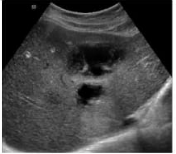
The sonographic image shows a complex fluid collection within the liver parenchyma, with internal echoes and possibly septations, consistent with an abscess. In the clinical context of post-procedural fever following a liver biopsy, a liver abscess is the most likely diagnosis.
A liver abscess appears on ultrasound as a hypoechoic or complex fluid collection that may contain internal debris, septations, or gas (which may produce reverberation artifacts). These features distinguish it from other post-procedural complications.
A cyst (Option A) typically appears as an anechoic, well-defined lesion with posterior acoustic enhancement and no internal debris---this does not match the image or clinical setting.
A biloma (Option B) is a bile collection that can appear similar to a cyst or fluid collection but typically occurs due to bile leak; however, fever and internal complexity on ultrasound more strongly suggest abscess.
A hematoma (Option D) may also appear complex but usually presents with pain and not fever unless secondarily infected. Over time, hematomas evolve in appearance but lack septations and gas unless superinfected.
Rumack, Carol M., et al. Diagnostic Ultrasound. 5th ed., Elsevier, 2018. Chapter: Hepatobiliary System, pp. 107--111.
American Institute of Ultrasound in Medicine (AIUM) Practice Parameter for the Performance of an Ultrasound Examination of the Abdomen and/or Retroperitoneum.