
 MENU
MENU
Which type of artifact is indicated by the arrows on this image?
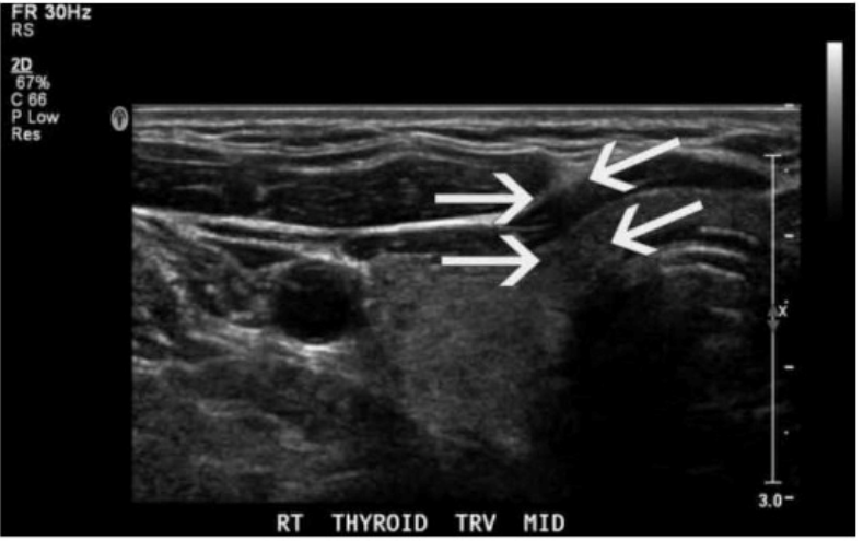
The ultrasound image of the thyroid clearly shows posterior shadowing originating from the lateral edges of a rounded structure, which is indicative of edge shadow artifact. Edge shadowing occurs when an ultrasound beam passes tangentially to a rounded or curved structure, such as a cyst or blood vessel. The difference in sound wave refraction and beam divergence at the edges leads to decreased echo signals deep to the edges, creating linear hypoechoic bands --- which is exactly what the arrows are pointing to in the image.
Edge shadow artifact is purely a result of beam physics and not a real anatomic or pathologic finding.
Key characteristics of edge shadowing:
Appears as a narrow, linear hypoechoic (dark) shadow extending deep to the edge of a curved interface (e.g., cyst, vessel, thyroid nodule)
Caused by refraction and beam deflection, leading to reduced beam intensity distal to the edges
Most commonly seen adjacent to cysts or fluid-filled structures
Differentiation from other options:
A . Focal enhancement: Appears as increased echogenicity distal to a fluid-filled structure due to lower attenuation of the sound beam through fluid (opposite of shadowing).
C . Speed error: A less common artifact that results in displacement of structures due to variation in assumed sound speed.
D . Comet tail: A reverberation artifact that appears as a series of closely spaced bright echoes, often associated with metallic objects or cholesterol crystals in adenomyomatosis.
Rumack CM, Wilson SR, Charboneau JW, Levine D. Diagnostic Ultrasound. 5th Edition. Elsevier, 2018. Chapter: Ultrasound Physics and Artifacts, pp. 38--42.
Kremkau FW. Sonography Principles and Instruments. 9th Edition. Elsevier, 2015. Chapter: Image Artifacts, pp. 132--136.